Advanced Dental Technology – Raleigh, NC
Delivering the Most Accurate Results
When it comes to repairing, improving, and rebuilding smiles, our team does not believe in cutting corners or providing mediocre results. Instead, we incorporate the use of the most advanced dental technologies available. From intraoral cameras to digital impressions and a CT/cone beam scanner, we ensure that our patients receive accurate results every time. Learn more about the equipment and solutions we use in our Raleigh dental practice.
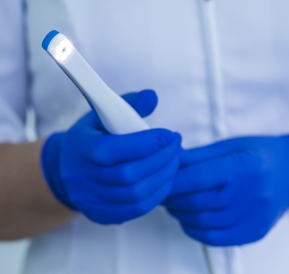
Digital Dental Impressions

Capturing a mold of a patient’s mouth is required when it comes to creating a customized restoration. Whether it is a crown, veneer, or a set of dentures, precise and accurate impressions are essential for a comfortable and reliable fit. Although messy dental putty is still used by many professionals, we opt for a more patient-friendly alternative – digital dental impressions. Using this handheld device, we can easily scan a person’s teeth and gums to create a three-dimensional model that is used by lab technicians to build a fully functional, natural-looking restoration.
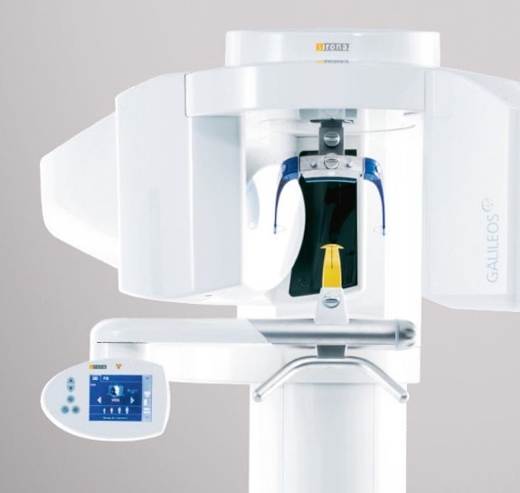
CT/Cone Beam Scanner

Complex dental treatments like dental implant placement, root canal therapy, and other similar procedures often require more enhanced imaging than those provided by X-rays. This is why we turn to the CT/Cone Beam Scanner. Showcasing a person’s teeth, bones, tissues, nerve pathways, blood vessels, and other nearby structures, we can formulate treatment plans more readily and thoroughly thanks to the high-resolution, 3D model that is produced with the help of hundreds of images captured by the 360-degree rotating arm.
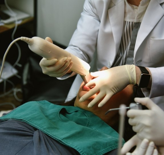
Intraoral Camera

The inner workings of a person’s mouth are no longer a mystery to a patient thanks to intraoral cameras. Instead of listening to the dentist explain what it is they see, an individual can now see in real-time what is developing inside their mouth. From small crevices of decay to more severe cases of gum disease, this camera-tipped, handheld device scans the mouth and projects the images onto a chairside monitor for easy viewing and more in-depth patient education.
